

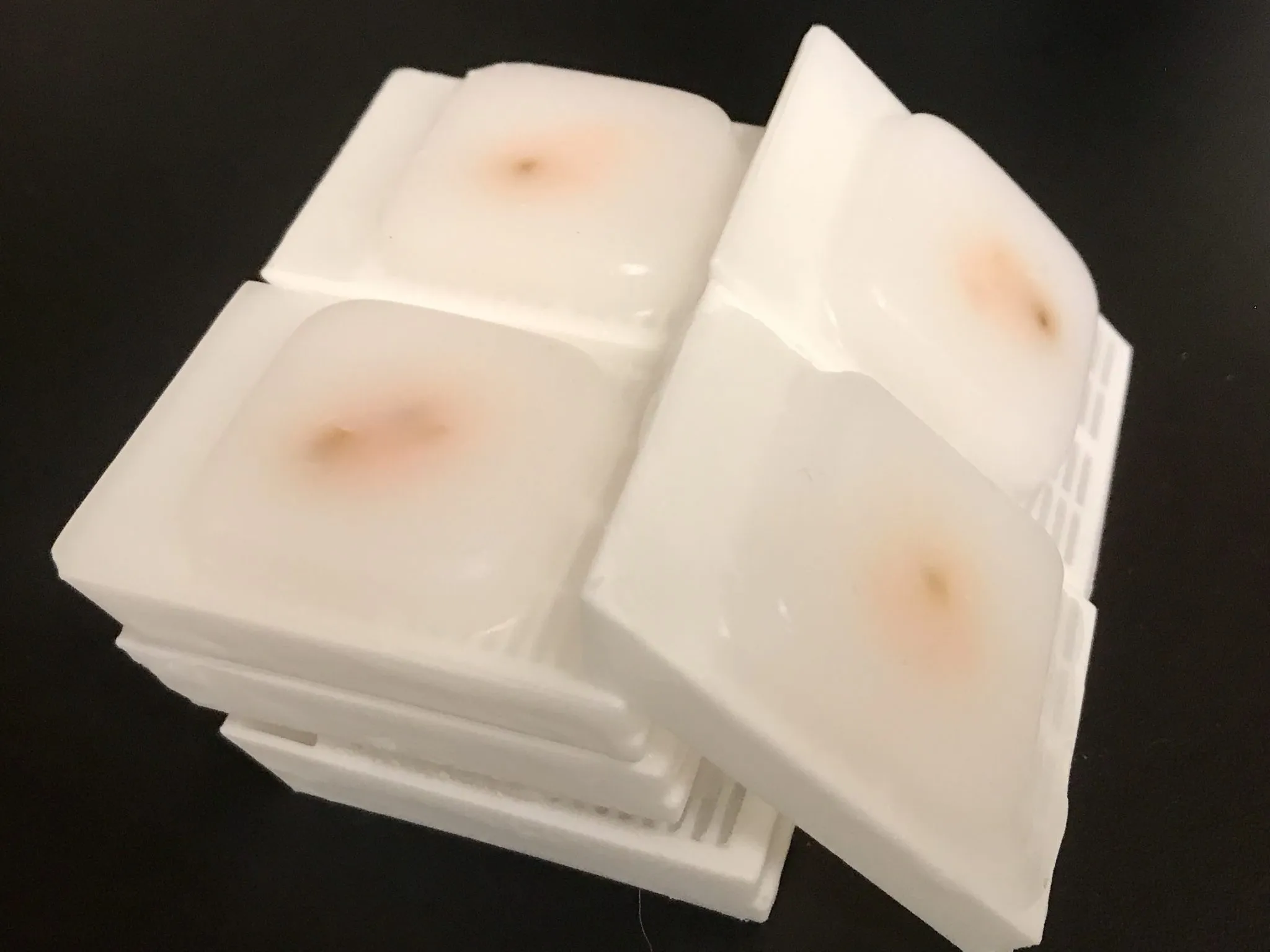
Archived tissue can be extremely valuable when researchers need to revisit an earlier study, compare historical samples, or explore a new biomarker without collecting fresh material. Properly stored ffpe blocks preserve tissue architecture well and can support many downstream histology and molecular pathology workflows.
The challenge is that preservation changes tissue chemistry. Formalin fixation creates cross-links that stabilize structure but can also hide protein epitopes. Researchers therefore need to balance preserved morphology with technical steps that restore antibody access before immunostaining.
Why FFPE Material Remains Valuable
Formalin-fixed, paraffin-embedded tissue is widely used because it provides durable specimens that can be sectioned repeatedly over time. Researchers can prepare new slides when a study expands, when additional markers are introduced, or when older findings need to be re-examined.
This flexibility makes archived tissue useful for longitudinal projects, retrospective research, and studies with limited specimen availability. Fixation history, storage conditions, and section age should still be documented because these factors may influence staining performance.
Recovering Hidden Protein Targets
A common difficulty in immunohistochemistry is reduced antibody access after formalin fixation. For many targets, heat-induced epitope retrieval with citrate buffer can help expose masked sites and improve antibody access before the staining procedure begins.
Cross-linking can otherwise produce weak or inconsistent signal when tissue is stained without appropriate pretreatment. Retrieval conditions should therefore be considered alongside the antibody, expected localization, tissue type, and fixation history.
Choose Retrieval Conditions Deliberately
Retrieval is not a universal one-setting step. Buffer composition, pH, temperature, heating time, and cooling conditions can change both signal intensity and tissue integrity. A protocol that performs well for one antibody may not suit another.
Researchers should optimize retrieval using suitable control tissue before applying it to a large study. This reduces the risk of consuming valuable sections while testing conditions that produce excessive background, weak signal, or structural damage.
Protect Section Quality Before Staining
Thin, uniform sections are important for reproducible immunostaining. Poor adhesion, incomplete deparaffinization, folds, tears, or uneven thickness can interfere with antibody penetration and make staining difficult to compare between samples.
Charged slides, controlled drying, and consistent sectioning can improve handling during retrieval and staining. Because heat retrieval can place stress on sections, good slide adhesion becomes especially important when archived specimens are fragile.
Use Controls to Separate Technique From Biology
Positive controls confirm that the antibody and detection system can produce the expected pattern under the chosen retrieval conditions. Negative controls help reveal nonspecific signal, reagent background, or unwanted binding.
Controls should be processed in the same run as research sections whenever possible. This makes it easier to decide whether an unexpected result reflects true biology, tissue quality, antibody behavior, or a technical change in the workflow.
Keep Antibody Optimization Separate
Retrieval is only one part of immunohistochemistry. Antibody concentration, incubation time, blocking strategy, detection chemistry, and counterstaining also influence the finished slide. Increasing antibody concentration should not automatically compensate for poor retrieval.
A better approach is to optimize variables systematically. Researchers can establish suitable retrieval first, then refine antibody dilution and detection conditions until the signal is specific, interpretable, and consistent across experimental samples.
Standardize Imaging and Scoring
Once staining is complete, image acquisition should remain controlled. Magnification, illumination, exposure, white balance, and digital processing can all change the apparent strength of a protein signal.
Scoring criteria should also be defined before the full slide set is reviewed. Researchers may evaluate positive-cell percentage, staining intensity, cellular localization, or region-specific patterns depending on the biological question and study design.
Plan Section Use Before Cutting
Archived blocks may contain limited remaining tissue, especially after years of previous research. A sectioning plan can allocate slides for H&E review, immunostaining, controls, repeat testing, and possible future assays without unnecessary recutting.
Recording section order is also useful when adjacent slides are compared. It allows researchers to match protein expression with nearby morphology while maintaining a clear record of how each portion of the specimen was used.
Conclusion
Preserved tissue can remain highly informative when researchers account for the effects of fixation and storage. Strong morphology is only part of the equation; successful protein localization also depends on exposing relevant epitopes without damaging the section.
By combining careful section preparation, optimized retrieval, appropriate controls, disciplined antibody testing, and consistent imaging, research teams can obtain meaningful protein information from archived specimens while using limited material efficiently.